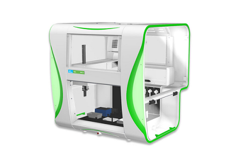
JANUS® G3 NGS Express™ Workstation
It uses an intuitive interface to guide users through protocol selection and deck setup
An optional enclosure helps to minimize sample contamination
The PKeye™ mobile operations monitor empowers full walk-away automation through the use of integrated deck cameras.
- Product description
- Citations
The JANUS® G3 NGS Express™ workstation is a benchtop liquid handler designed to simplify and reduce errors in low-to-moderate throughput next generation sequencing library construction. It uses an intuitive interface to guide users through protocol selection and deck setup, as well as, displays in-run status and details. An optional enclosure not only helps to minimize sample contamination, but the easy to read status lights provide visual feedback for true walkaway sample preparation. Another advanced option is the PKeye™ mobile operations monitor, which empowers full walk-away automation through the use of integrated deck cameras.
NGS applications automated on the JANUS G3 NGS Express workstation include:
Library preparation
Amplicon preparation
Target capture preparation
Sample normalization
Specifications
Precision Automated Pipetting with a Wide Dynamic Volume Range
| Pipetting Arm | Volume | % CV | Tip/Head | Condition |
| Varispan | 5 µL | < 2% | 20 µL disposable tip | Distilled water, 500 µL syringe |
| Varispan | 50 µL | < 1% | Fixed tip | Distilled water, 500 µL syring |
Microplate Deck Capacity: 12
| Dimensions | |
| JANUS G3 NGS Express workstation without an enclosure | |
| Height | 864 mm | 34 in |
| Width | 788 mm | 31 in |
| Depth | 838 mm | 33 in |
| JANUS G3 NGS Express workstation with an enclosure | |
| Height | 965 mm | 38 in |
| Width | 1016 mm | 40 in |
| Depth | 890 mm | 35 in |
Publications that Cite Using the Custom JANUS® G3 NGS Express™ Workstation
Bagheri, H., Friedman, H., Shao, H., Chong, Y., Lo, C., Emran, F., . . . Peterson, A. (2018). TIE: A Method to Electroporate Long DNA Templates into Preimplantation Embryos for CRISPR-Cas9 Gene Editing. The CRISPR Journal, 1(3), 223-229. doi:10.1089/crispr.2017.0020.
Briem, F., Zeisler, C., Guenay, Y., Staudacher, K., Vogt, H., & Traugott, M. (2018). Identifying plant DNA in the sponging–feeding insect pest Drosophila suzukii. Journal of Pest Science, 91(3), 985-994. doi:10.1007/s10340-018-0963-3.
Carpenter, S. L., et al. (2014) Use of Dried Blood Spots for High through-Put, Rapid Turnaround Mutational Analysis in Patients with Hemophilia, Blood, 124(5034).
Lee, J., Wang, J., Sa, J. K., Ladewig, E., Lee, H., Lee, I., . . . Nam, D. (2017). Spatiotemporal genomic architecture informs precision oncology in glioblastoma. Nature Genetics,49(4), 594-599. doi:10.1038/ng.3806
Murra, M., Lützen, L., Barut, A., Zbinden, R., Lund, M., Villesen, P., & Nørskov-Lauritsen, N. (2018). Whole-Genome Sequencing of Aggregatibacter Species Isolated from Human Clinical Specimens and Description of Aggregatibacter kilianii sp. nov. Journal of Clinical Microbiology,56(7). doi:10.1128/jcm.00053-18.
Xu, L., Brito, I. L., Alm, E. J., & Blainey, P. C. (2016). Virtual microfluidics for digital quantification and single-cell sequencing. Nature Methods,13(9), 759-762. doi:10.1038/nmeth.3955.


